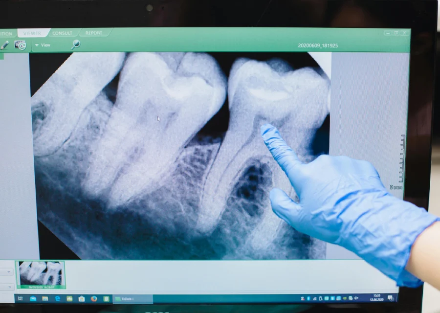
What Is an IOPA X-Ray and Why Is It Important in Dentistry?
Dental health often involves a close examination of the teeth, gums and surrounding structures to diagnose and treat various conditions. One of the essential diagnostic tools in dentistry is the IOPA X-ray, which stands for Intraoral Periapical X-ray. This specialized imaging technique provides a detailed view of specific areas within the oral cavity, making it an invaluable tool for both routine checkups and complex dental procedures.
This article delves into what an IOPA X-ray is, how it works and why it is a critical component of modern dental care.
An IOPA X-ray (Intraoral Periapical X-ray) is a type of dental imaging that focuses on a small section of the mouth. Unlike panoramic X-rays that capture the entire mouth, IOPA X-rays provide highly detailed images of a specific tooth or a group of teeth, including the root, surrounding bone and nearby structures.
Key Features of IOPA X-Rays
-
High Precision: Captures fine details of the tooth structure and adjacent areas.
-
Compact Imaging: Focuses on a limited section of the mouth for targeted analysis.
-
Real-Time Diagnostics: Offers immediate insights for accurate diagnosis and treatment planning.
IOPA X-rays use low doses of radiation to penetrate oral tissues and create detailed images. The process involves placing a small sensor or film inside the patient’s mouth and directing a controlled X-ray beam to the target area.
Steps in the Process
-
Positioning the Sensor: The dentist or technician positions the sensor near the targeted tooth.
-
Directing the X-ray Beam: A handheld or mounted X-ray device emits the beam toward the sensor.
-
Capturing the Image: The sensor captures the X-rays and transmits the image to a computer or film for analysis.
Safety Measures
-
Use of lead aprons to protect the patient’s body.
-
Low radiation exposure ensures safety without compromising image quality.
Why Is an IOPA X-Ray Important in Dentistry?
IOPA X-rays play a vital role in diagnosing and managing dental conditions effectively. Their high level of detail enables dentists to detect issues that may not be visible during a standard oral examination.
Benefits of IOPA X-Rays
1. Accurate Diagnosis of Dental Problems
-
Identifies cavities, infections and fractures with precision.
-
Detects root canal infections and abscesses.
-
Monitors the health of the bone surrounding the tooth.
2. Essential for Root Canal Treatment
-
Helps locate the exact position of tooth roots and canals.
-
Guides the dentist in cleaning and filling the root canal system.
3. Supports Dental Surgeries
-
Provides a clear view for procedures like tooth extractions or implant placement.
-
Assesses bone density and structure for surgical planning.
4. Monitoring Dental Restorations
-
Evaluates the fit and condition of fillings, crowns and bridges.
-
Detects potential complications early, ensuring longevity of restorations.
When Do Dentists Recommend an IOPA X-Ray?
Dentists may recommend IOPA X-rays for a variety of reasons, depending on the patient’s symptoms and treatment needs.
Common Scenarios
-
Persistent Tooth Pain: To identify hidden cavities, cracks or infections.
-
Post-Treatment Monitoring: To ensure proper healing after a root canal or extraction.
-
Orthodontic Assessment: To evaluate tooth alignment and root positioning.
-
Periodontal Evaluation: To assess bone loss or gum disease severity.
-
Pre-Surgical Planning: For precise mapping of structures before dental surgery.
Advantages of Using IOPA X-Rays
The IOPA X-ray stands out for its precision and utility in dental diagnostics. Here are its primary advantages:
1. Detailed Imaging
IOPA X-rays capture intricate details of tooth roots, bone levels and surrounding structures, allowing for accurate diagnosis.
2. Minimally Invasive
The procedure is quick, painless and requires minimal preparation.
3. Immediate Results
Digital IOPA X-rays provide instant images, reducing wait times for diagnosis and treatment planning.
4. Cost-Effective
As a focused imaging technique, IOPA X-rays are often more affordable compared to full-mouth scans.
Precautions and Safety
Although IOPA X-rays involve radiation, modern techniques minimize exposure, making them safe for most patients. However, certain precautions should be observed:
Safety Guidelines
-
Pregnant Patients: Inform your dentist if you are pregnant; alternative imaging methods may be considered.
-
Protective Gear: Always wear the provided lead apron and thyroid collar.
-
Frequency: Limit the number of X-rays to what is clinically necessary.
Final Thoughts
An IOPA X-ray is a cornerstone of modern dental diagnostics, offering unparalleled precision and utility in detecting and managing oral health issues. Whether it’s identifying a root canal infection, planning dental surgery or monitoring restorations, this imaging technique ensures accurate and efficient care. By understanding the importance of IOPA X-rays, patients can feel confident about their role in achieving optimal oral health.
If your dentist recommends an IOPA X-ray, rest assured that it’s a safe, effective tool to support your dental health journey.
To schedule an appointment at ‘Sukumar Dental Clinic’ call +91-7418210108 or WhatsApp Dr. Sukumar at +91-9655225002. We take pride in having the top dental clinic in Palayamkottai, Tirunelveli. Alternatively, you can email us at info@sukumardental.com


