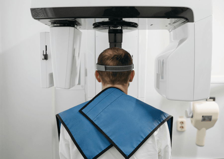
Dental imaging has come a long way from traditional X-rays to the advanced technology of Cone Beam Computed Tomography (CBCT). CBCT is transforming the way dentists diagnose and treat dental issues, offering a more accurate and detailed view of a patient’s oral health. In this post, we’ll explore how CBCT works and why it’s becoming the go-to tool for dental imaging, revolutionizing dental care in many ways.
What is CBCT?
Cone Beam Computed Tomography (CBCT) is a specialized type of X-ray technology that captures 3D images of the teeth, bone sand soft tissues in the mouth. Unlike traditional 2D X-rays, CBCT provides a three-dimensional view, which offers much more detailed information. The term “cone beam” refers to the cone-shaped X-ray beam used in the scanning process, which helps capture comprehensive images in a short amount of time.
CBCT is used in various dental procedures, including implants, root canals, orthodontics and even the diagnosis of diseases or injuries affecting the mouth and jaw.
How Does CBCT Work?
CBCT uses a rotating X-ray beam that circles around the patient’s head while a special detector captures images from all angles. These images are then reconstructed by a computer into a 3D image that shows a detailed view of the patient’s mouth, teeth, jawbone and even sinuses.
This process is much more detailed than traditional X-rays, which only provide a 2D image of the area being scanned. The 3D images produced by CBCT are incredibly helpful for dentists in planning treatments and diagnosing issues with higher accuracy.
Key Advantages of CBCT Over Traditional X-Rays
- Detailed 3D Imaging
One of the biggest advantages of CBCT over traditional X-rays is the ability to create three-dimensional images. Traditional X-rays, such as bitewings or panoramic X-rays, only provide a flat, 2D image of the teeth and jaw. This can sometimes make it difficult to get a full picture of the underlying issues, such as cavities, bone loss or root infections.
With CBCT, dentists can examine the patient’s mouth from every angle, which leads to a more comprehensive understanding of the patient’s dental health. This 3D capability allows dentists to view structures in a way that was not possible with traditional X-rays, helping them catch issues that might otherwise go unnoticed.
- Better Accuracy for Treatment Planning
The precision of CBCT imaging makes it especially valuable for procedures that require careful planning, such as dental implants. Before placing an implant, it’s important to assess the density and shape of the bone to ensure there’s enough support for the implant. With traditional 2D X-rays, it’s difficult to assess bone structures accurately because they provide only a single plane of view.
With CBCT, dentists can evaluate the 3D shape and density of the bone in all directions. This information is crucial for placing implants precisely in the best location, reducing the chances of complications and improving the long-term success of the implant.
- Improved Diagnosis of Dental Issues
Traditional X-rays are great for identifying issues like cavities, infections and fractures, but they can miss certain problems, especially those hidden beneath the surface. CBCT provides a more detailed and clearer picture, allowing dentists to detect issues that might not show up in a 2D image. This includes things like small fractures, bone loss, tumors, cysts and other abnormalities in the jaw and facial bones.
CBCT is particularly helpful for diagnosing conditions like TMJ (temporomandibular joint) disorders, as it provides a better view of the jaw’s movement and alignment. This allows for a more accurate diagnosis and the development of a tailored treatment plan.
- Lower Radiation Exposure
While CBCT does use X-rays, it often uses much less radiation than traditional CT scans, which is an important safety factor for both patients and dentists. Traditional dental X-rays generally expose the patient to a small amount of radiation, but a full CT scan can involve a much higher dose. In contrast, CBCT scans are designed to provide detailed images with lower radiation, making them a safer option for dental imaging.
Additionally, the shorter time required for a CBCT scan means less overall radiation exposure. Many CBCT machines can deliver the necessary images in a matter of seconds, further minimizing the risk of radiation.
- Faster and More Efficient
Traditional X-rays require multiple images to be taken from different angles to get a full picture of a patient’s dental health. For example, bitewing X-rays might only show the teeth, while panoramic X-rays provide a wider view of the mouth and jaw. With CBCT, all of this can be captured in one scan.
The speed of the CBCT scan is another benefit. It typically takes just a few seconds to complete, compared to the longer process of taking and developing multiple traditional X-rays. This efficiency is especially helpful in emergency situations, where quick imaging is needed to assess injuries or diagnose conditions.
- Better for Complex Cases
For more complicated dental issues, such as impacted teeth, abnormal tooth development or serious infections, CBCT provides a level of detail that traditional X-rays simply can’t match. It’s also invaluable for cases that involve oral surgery, as it allows the surgeon to plan the procedure with a precise understanding of the patient’s anatomy.
For instance, in cases where a patient needs a root canal, CBCT can help visualize the roots of the tooth in 3D, revealing any hidden infections or cracks that might not show up on traditional X-rays. This leads to more accurate treatment and better outcomes.
- Helps in Orthodontics
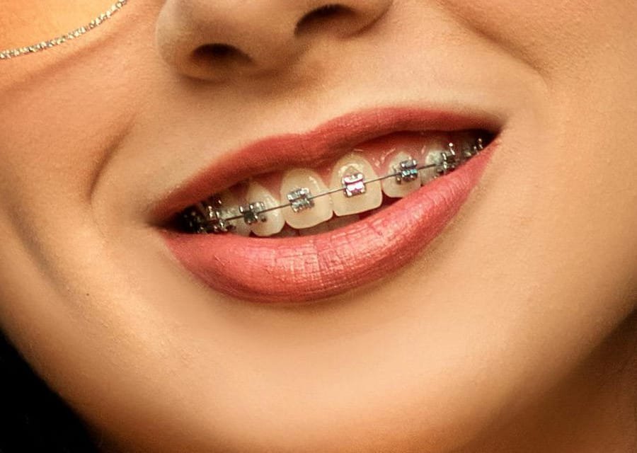
CBCT is also a game-changer for orthodontics. While traditional X-rays can show basic details about the alignment of teeth, CBCT allows orthodontists to assess bone structure, tooth positions and even airways in a 3D format. This is particularly useful in planning complex orthodontic treatments, including the treatment of jaw alignment and sleep apnea issues.
Conclusion
CBCT is revolutionizing dental imaging by offering detailed 3D images, improving diagnostic accuracy and making treatment planning more precise. Its ability to provide comprehensive views of the teeth, bones and surrounding tissues makes it a powerful tool for both routine dental care and complex procedures like implants, root canals and orthodontics.
While traditional X-rays are still widely used and effective for many dental issues, CBCT is quickly becoming the preferred choice for dentists who want to ensure the best possible care for their patients. With its lower radiation exposure, faster scan times and enhanced accuracy, CBCT is truly changing the landscape of dental imaging, leading to better outcomes and more efficient treatments for patients.
To schedule an appointment at ‘Sukumar Dental Clinic’ call +91-7418210108 or WhatsApp Dr. Sukumar at +91-9655225002. We take pride in having the top dental clinic in Palayamkottai, Tirunelveli. Alternatively, you can email us at info@sukumardental.com


